Bedankt voor het vertrouwen het afgelopen jaar! Om jou te bedanken bieden we GRATIS verzending (in België) aan op alles gedurende de hele maand januari.
- Afhalen na 1 uur in een winkel met voorraad
- In januari gratis thuislevering in België
- Ruim aanbod met 7 miljoen producten
Bedankt voor het vertrouwen het afgelopen jaar! Om jou te bedanken bieden we GRATIS verzending (in België) aan op alles gedurende de hele maand januari.
- Afhalen na 1 uur in een winkel met voorraad
- In januari gratis thuislevering in België
- Ruim aanbod met 7 miljoen producten
Zoeken
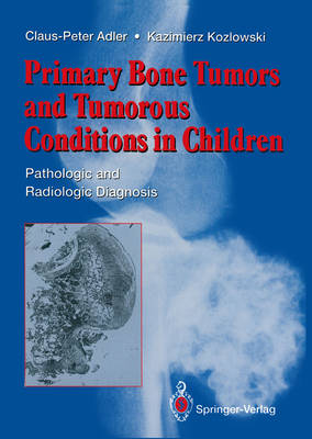
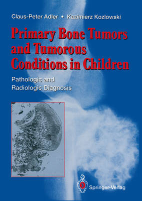
Primary Bone Tumors and Tumorous Conditions in Children
Pathologic and Radiologic Diagnosis
Claus-Peter Adler, Kazimierz Kozlowski
Paperback
€ 139,95
+ 279 punten
Omschrijving
We welcome the publication of this volume, which discusses the diagnosis of bone tumours with particular reference to children and adolescents. However, bone tumors often show an extreme variety of structures which confuse even experienced bone pathologists.
Specificaties
Betrokkenen
- Auteur(s):
- Uitgeverij:
Inhoud
- Aantal bladzijden:
- 267
Eigenschappen
- Productcode (EAN):
- 9781447119531
- Verschijningsdatum:
- 20/11/2011
- Uitvoering:
- Paperback
- Afmetingen:
- 193 mm x 270 mm
- Gewicht:
- 622 g

Alleen bij Standaard Boekhandel
+ 279 punten op je klantenkaart van Standaard Boekhandel
Beoordelingen
We publiceren alleen reviews die voldoen aan de voorwaarden voor reviews. Bekijk onze voorwaarden voor reviews.


