Bedankt voor het vertrouwen het afgelopen jaar! Om jou te bedanken bieden we GRATIS verzending (in België) aan op alles gedurende de hele maand januari.
- Afhalen na 1 uur in een winkel met voorraad
- In januari gratis thuislevering in België
- Ruim aanbod met 7 miljoen producten
Bedankt voor het vertrouwen het afgelopen jaar! Om jou te bedanken bieden we GRATIS verzending (in België) aan op alles gedurende de hele maand januari.
- Afhalen na 1 uur in een winkel met voorraad
- In januari gratis thuislevering in België
- Ruim aanbod met 7 miljoen producten
Zoeken
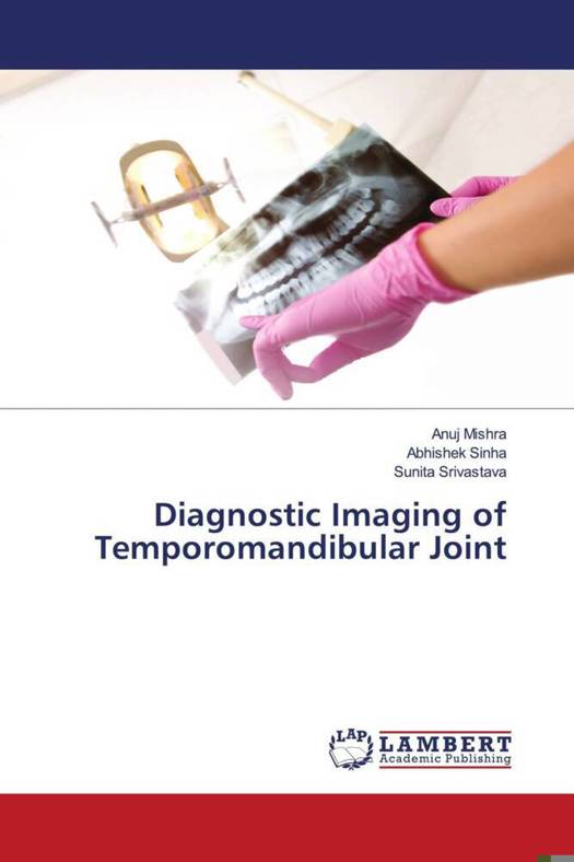
Diagnostic Imaging of Temporomandibular Joint
Anuj Mishra, Abhishek Sinha, Sunita Srivastava
Paperback | Engels
€ 55,95
+ 111 punten
Omschrijving
After a thorough review of the literature, it is clear that multiple imaging modalities are available for evaluation of the TMJ. The need for TMJ imaging should be assessed on an individual basis, depending on the signs and symptoms obtained and the working diagnosis. An Orthopantomogram within the framework of the "joint program" helps us to discover small bone defects or pathological positions of the joint condyles. However, the Arthography is an invasive examination and is accompanied by the risk of allergic reaction connected with contrast substance application. The possibility of imaging soft tissues close to the joint and the possibility of "3D" reconstruction of bone structures are a great advantage. Based on the evidence currently available, MRI continues to be the gold standard for imaging disk position and the soft tissues of the TMJ, including joint effusions. In contrast, CT is the ideal imaging choice for evaluating hard tissues, adding improvement in accessibility and radiation dosage with the use of the new cone-beam CT. Ultrasonography, as a completely non-invasive procedure, in diagnosing functional temporomandibular defects. Its great advantage mainly consists
Specificaties
Betrokkenen
- Auteur(s):
- Uitgeverij:
Inhoud
- Aantal bladzijden:
- 100
- Taal:
- Engels
Eigenschappen
- Productcode (EAN):
- 9783659767395
- Verschijningsdatum:
- 21/08/2015
- Uitvoering:
- Paperback
- Afmetingen:
- 150 mm x 220 mm
- Gewicht:
- 159 g

Alleen bij Standaard Boekhandel
+ 111 punten op je klantenkaart van Standaard Boekhandel
Beoordelingen
We publiceren alleen reviews die voldoen aan de voorwaarden voor reviews. Bekijk onze voorwaarden voor reviews.


