- Afhalen na 1 uur in een winkel met voorraad
- Gratis thuislevering in België vanaf € 30
- Ruim aanbod met 7 miljoen producten
- Afhalen na 1 uur in een winkel met voorraad
- Gratis thuislevering in België vanaf € 30
- Ruim aanbod met 7 miljoen producten
Zoeken
€ 80,95
+ 161 punten
Omschrijving
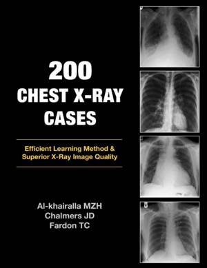
Modern medical practice has seen many advances in imaging over the past ten years. Magnetic Resonance Imaging, CT scanning and Ultrasound investigations have all been added to the repertoire of normal practice. However, the humble chest X-ray remains a crucial first line investigation - particularly for acute medical admissions. Many chest X-rays are requested for a specific purpose e.g. confirmation of pneumonia, but the image may reveal features of previously unsuspected disease of another body system. Digital storage of X-ray images means that a chest X-ray may be viewed at any computer workstation in your hospital. Clinicians now have the opportunity to view these images without waiting for the X-ray packet to be delivered. This can only be advantageous for the patient if the clinician knows what to look for on the image. This book takes you through 200 images in a stimulating manner designed to improve your confidence in reporting the humble chest X-ray.
Specificaties
Betrokkenen
- Auteur(s):
- Uitgeverij:
Inhoud
- Aantal bladzijden:
- 216
- Taal:
- Engels
Eigenschappen
- Productcode (EAN):
- 9781905006366
- Verschijningsdatum:
- 21/10/2009
- Uitvoering:
- Paperback
- Formaat:
- Trade paperback (VS)
- Afmetingen:
- 216 mm x 279 mm
- Gewicht:
- 512 g

Alleen bij Standaard Boekhandel
+ 161 punten op je klantenkaart van Standaard Boekhandel
Beoordelingen
We publiceren alleen reviews die voldoen aan de voorwaarden voor reviews. Bekijk onze voorwaarden voor reviews.


