
Je cadeautjes zeker op tijd in huis hebben voor de feestdagen? Kom langs in onze winkels en vind het perfecte geschenk!
- Afhalen na 1 uur in een winkel met voorraad
- Gratis thuislevering in België vanaf € 30
- Ruim aanbod met 7 miljoen producten
Je cadeautjes zeker op tijd in huis hebben voor de feestdagen? Kom langs in onze winkels en vind het perfecte geschenk!
- Afhalen na 1 uur in een winkel met voorraad
- Gratis thuislevering in België vanaf € 30
- Ruim aanbod met 7 miljoen producten


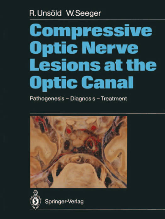
Compressive Optic Nerve Lesions at the Optic Canal
Pathogenesis - Diagnosis - Treatment
Renate Unsöld, Wolfgang Seeger
Paperback | Engels
€ 147,95
+ 295 punten
Omschrijving
The optic canal, in particular its intracranial end, represents a "locus minoris resistentiae" for optic nerve compression in a variety of pathologic conditions. The intracranial optic nerve shares the limited space within this narrow passage with the carotid and ophthalmic artery, all being surrounded by bone and rigid dura. Any pathological condition going along with an increase of soft tissue volume, such as in optic nerve sheath tumors, parasellar neoplasms, dolichoectasia of the carotid and/ or ophthalmic artery, hematomas, etc., or reduction of the lumen of the bony optic canal by hyperpneumatization of the sphenoid sinus, hyperostosis or developmental abnormalities must act as a space-occupying lesion causing optic nerve compression either by pressing the nerve against the vessel or the neighboring dura or bone. The spectrum of clinical signs and symptoms of optic nerve compression in this area is rather wide and includes acute as well as slowly progressive visual loss and all kinds of visual field defects in the presence of a normal disk, papilledema, pri mary optic atrophy or cavernous optic atrophy mimicking var ious clinical disease entities such as retrobulbar optic neuritis, anterior and posterior ischemic optic neuropathy, soft glaucoma and others. Some of the lesions causing optic nerve compression in this area are rather small and need to be visualized or excluded by thin section CT such as pneumosinus dilatans of the sphenoid bone, dolichoectasia of the internal carotid artery, small men ingiomas around the optic foramen and others."
Specificaties
Betrokkenen
- Auteur(s):
- Uitgeverij:
Inhoud
- Aantal bladzijden:
- 140
- Taal:
- Engels
Eigenschappen
- Productcode (EAN):
- 9783642733840
- Verschijningsdatum:
- 6/12/2011
- Uitvoering:
- Paperback
- Formaat:
- Trade paperback (VS)
- Afmetingen:
- 210 mm x 279 mm
- Gewicht:
- 367 g


Alleen bij Standaard Boekhandel
+ 295 punten op je klantenkaart van Standaard Boekhandel
CADEAU


Beoordelingen
We publiceren alleen reviews die voldoen aan de voorwaarden voor reviews. Bekijk onze voorwaarden voor reviews.