- Afhalen na 1 uur in een winkel met voorraad
- Gratis thuislevering in België vanaf € 30
- Ruim aanbod met 7 miljoen producten
- Afhalen na 1 uur in een winkel met voorraad
- Gratis thuislevering in België vanaf € 30
- Ruim aanbod met 7 miljoen producten

€ 261,95
+ 523 punten
Omschrijving
Each volume in this richly illustrated series, published in association with the Papanicolaou Society of Cytopathology, provides an organ-based approach to the cytologic and histologic diagnosis of small tissue samples. Benign, pre-malignant and malignant entities are presented in a well-organized and standardized format, with high-resolution color photomicrographs, tables, and lists of key specific morphologic criteria. Example vignettes allow the reader to assimilate the diagnostic principles in a case-based format. This volume provides comprehensive coverage of both surgical pathology and cytopathology of focal liver lesions. Extensively illustrated throughout, it contains key cytologic and histologic features, practical points, radiologic and morphologic pictures, flow charts, and tabulated summaries for easy comprehensive overview and quick reference and provides a pragmatic algorithmic approach to cytohistologic diagnosis. With over 700 printed photomicrographs and a CD-ROM offering all images in a downloadable format, this is an important resource for all anatomic pathologists.
Specificaties
Betrokkenen
- Auteur(s):
- Uitgeverij:
Inhoud
- Aantal bladzijden:
- 307
- Taal:
- Engels
- Reeks:
Eigenschappen
- Productcode (EAN):
- 9781107024175
- Verschijningsdatum:
- 6/03/2014
- Uitvoering:
- Boek
- Afmetingen:
- 221 mm x 279 mm
- Gewicht:
- 1338 g


Alleen bij Standaard Boekhandel
+ 523 punten op je klantenkaart van Standaard Boekhandel
Cadeau

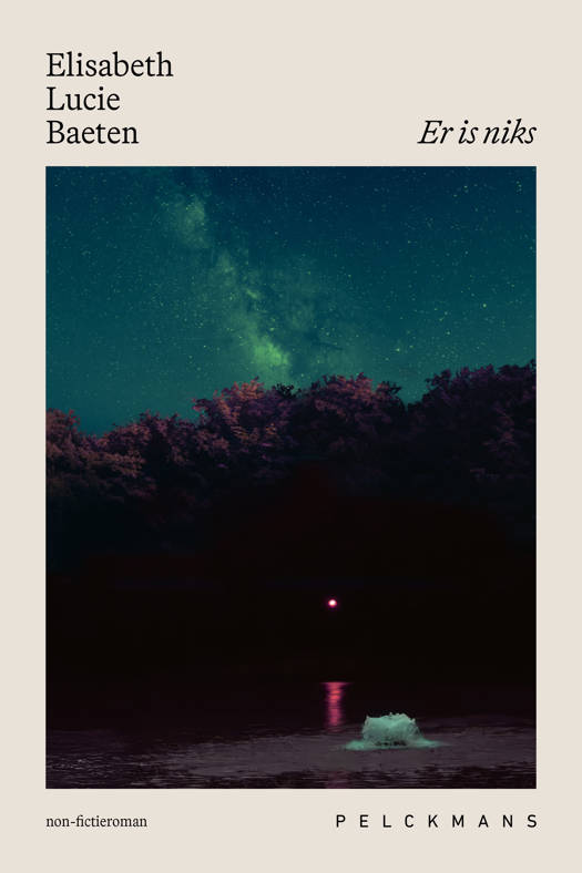
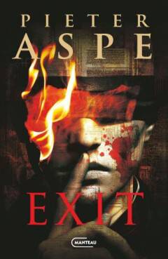
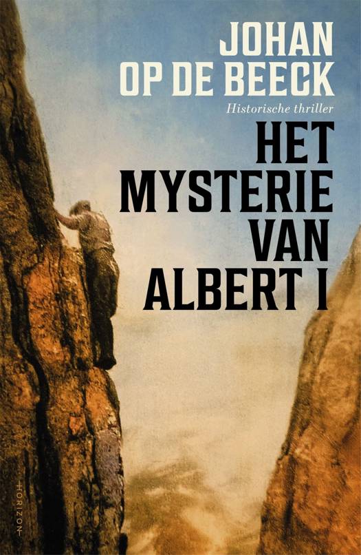
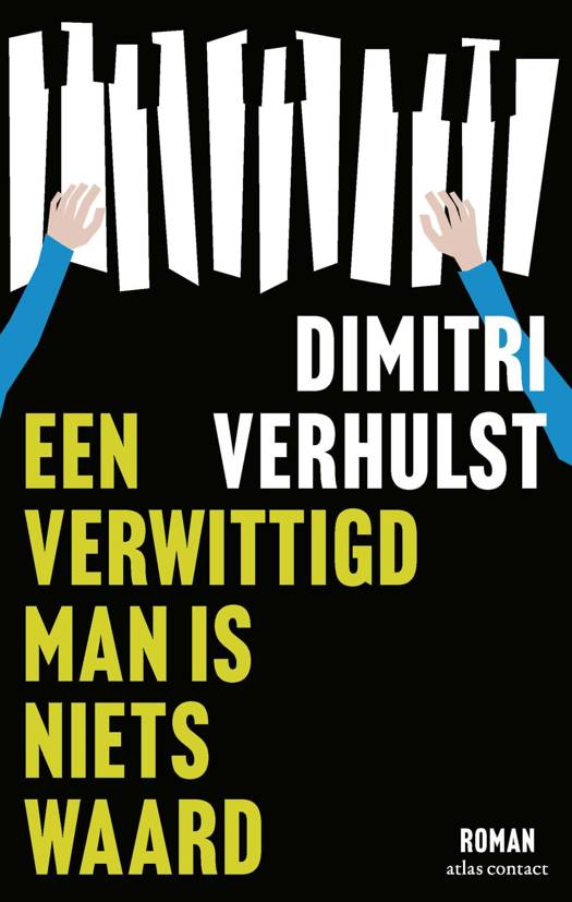
AANGERADEN


Beoordelingen
We publiceren alleen reviews die voldoen aan de voorwaarden voor reviews. Bekijk onze voorwaarden voor reviews.